Manuscript accepted on : 05 February 2012
Published online on: --
In vitro Conservation Approach of Rare Medicinal Plant Ceropegia juncea Roxb. (Asclepiadaceae)
M. N. Abubacker1* and G. Dheepan2
1Department of Biotechnology, National College, Tiruchirappalli - 620 001, India.
2Department of Botany, National College, Tiruchirappalli - 620 001, India.
ABSTRACT: In vitro conservation protocol for rare medicinal plant Ceropegia juncea was developed using nodal explants by culturing on Murashige and Skoog (MS) medium. The maximum number of greenish nodular callus was induced in MS + 6-benzylaminopurine (BAP) 1.5 mg/l + 2,4-dichlorophenoxy acetic acid (2,4-D) 1.5 mg/l and massive callus induction was in MS + Indole-3-Butyric Acid (IBA) 1.0 mg/l + BAP 1.0 mg/l + 2,4-D 0.5 mg/l. The greenish nodular calli were raised on the MS medium supplemented with BAP 3.0 mg/l + kinetine (Kin) 0.5 mg/l + Indole-3-Butyric Acid (IBA) 0.5 mg/l + Naphthalene Acetic Acid (NAA) 0.5 mg/l for shoot induction. Root induction was achieved in MS + Indole-3-Acetic Acid (IAA) 0.5 mg/l + NAA 0.5 mg/l + IBA 0.5 mg/l + BAP 1.0 mg/l. The plantlets were established, acclimatized and thrived in greenhouse and then in natural environmental condition. The in vitro conservation protocol developed in this study provides a basis for germplasm conservation of this medicinal plant.
KEYWORDS: Callus; Ceropegia juncea; in vitro conservation; MS medium
Download this article as:| Copy the following to cite this article: Abubacker M. N, Dheepan G. In vitro Conservation Approach of Rare Medicinal Plant Ceropegia juncea Roxb. (Asclepiadaceae). Biosci Biotech Res Asia 2012;9(1) |
| Copy the following to cite this URL: Abubacker M. N, Dheepan G. In vitro Conservation Approach of Rare Medicinal Plant Ceropegia juncea Roxb. (Asclepiadaceae). Biosci Biotech Res Asia 2012;9(1). Available from: https://www.biotech-asia.org/?p=9713 |
Introduction
India has a rich and varied heritage of biodiversity, encompassing a wide spectrum of habitats from rasin forests to alpine vegetation and from temperate forests to costal wetlands. In India, about 1500 rare and threatened species of both flowering and non-flowering plant groups were identified during the last two decades. The threat to a majority of them could be attributed to anthropogenic factors like habitat destruction due to grazing, urbanization and other developmental activities and exploitation (Myers, 1988).
Ceropegia L. is an old world tropical genus containing about 200 species of which 48 Ceropegia species found in India (Bruyns, 2003). Twenty eight species of Ceropegia are endemic to the Peninsular India (Ansari, 1984 Ahmedulla and Nayar, 1986). The existence of the Ceropegia species has become restricted to remote pockets in the Himalayas and the Western Ghats, the two biodiversity hot spots. Ceropegia genus has now been placed under the category of rare and endangered plants (Nayar and Sastry, 1987; Chaturvedi et al., 1996; Jagtap et al., 2004)
Ceropegia juncea Roxb. (Asclepiadaceae) is an important medicinal herb, which is used as a source of ‘soma’ a plant drug of the Ayurvedic medicine with a wide variety of uses. The fleshy stem and root tubers used as a raw material for traditional and folk medicines for the treatments of stomach and gastric disorders (Nikam and Savant, 2009). C. bulbosa and C. tuberosa root tubers contain an alkaloid called Ceropegin (Nadkarni, 1976), consumed after cooking (Mabberley, 1997). The root tubers also contain starch, sugars, albuminoids, fats, gum, crude fiber and valuable constituents in many traditional Indian Ayurvedic drug preparations that are active against many diseases especially diarrhea and dysentery. The ceropegin is an analgesic drug, tranquilizer and acts against ulcers and inflammation (Adibatti et al., 1991). It is important to prevent the extinction of C. juncea for its taxonomic and ethnobotanical values. The present study was aimed at in vitro conservation approach of this rare medicinal plant.
Materials and Methods
Plant material and explants preparation
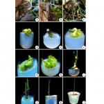
The plants of C. juncea were collected from a naturalized population in Nartharmalai (Pudukottai District), Tamil Nadu, India (Figure 1, a,b,c). For periodic harvest of explants, stocks were maintained in the herbal garden at Department of Botany, National College, Tiruchirappalli, Tamil Nadu. Nodal portions of succulent stem were tested for culture initiation. The young succulent stems were excised from the plants and kept under tap water with Tween 20 (4-5 drops) for 20 min. Nodal segments (1-2 cm long) was cut from these stems and was treated with fungicide Bavistin (0.5-1.0% w/v) and antibiotic streptomycin (0.05-0.1% w/v) for 10 min each. Each time the nodal segments were rinsed thrice in distilled water and then they were surface sterilized with aqueous solution of HgCl2 (0.1% w/v) for 2-3 min and washed thrice with sterilized distilled water. The explants were cut gently with a sterilized blade and explants were inoculated in culture tubes with modified MS media under aseptic conditions.
Culture medium and conditions
Basal Murashige and Skoog medium (Murashige and Skoog, 1962). (MS medium) was prepared using Hi media and Sigma chemicals. Sucrose (30% w/v) was added to medium and pH of medium was adjusted to 5.8 before adding agar-agar (8% w/v). Different growth regulators (BAP, 2, 4-D, IBA, Kin, NAA and IAA) at different concentrations and combinations were added to the medium. In the present study, the media were autoclaved at 121° C and 15 lbs pressure for 20 min. All the in vitro cultures were maintained at 24 ± 2° C and illuminated for 16 h with fluorescent light (18-24 m mol/m/sec) followed by 8 h dark period and the relative humidity was about 60-80% within the 25´150 mm culture tubes covered with cotton plug.
Callus, shoot and root induction and acclimatization
The healthy and normal nodal segment explants were inoculated in MS medium with different combinations of growth regulators. When the growth regulators failed to induce a specific response in callus, direct organogenesis and multiple shoot production at the end of the first cycle, it was marked as inappropriate combination. Twenty cultures were raised for each treatment and all experiments were repeated thrice. BAP, 2, 4-D, IAA and IBA combinations were tested to estimate the callus production. BAP, Kin, IAA, IBA and NAA combinations were used for shoot induction. The regenerated shoots were inoculated for rooting medium supplemented with IAA, IBA, NAA and BAP combinations. The rooted shoots were washed with sterile distilled water to remove the traces of medium. The in vitro rooted plantlets were transferred to cups containing autoclaved sand. Those cups were covered initially with polythene bags to maintain humidity and placed in mist chamber. After every alternative day, quarter strength MS medium salt solution was supplied to the plantlets. After two weeks of growth, the complete plants were established, acclimatized and thrived in green house condition. The acclimatized plants after two weeks were then transferred to natural environment and established into healthy plants (Figure 1, l)
Statistical analysis
All statistical analyses were performed at 0.05% level, using the statistical software (SPSS inc. Chicago; USA).
Results and Discussion
The combination of BAP, IBA and 2,4-D induced an excellent amount of massive callus from the nodal segments of Ceropegia juncea and the morphology of the callus was friable, yellowish green and greenish in colour and nodular in its nature (Table 1).
Table 1 – Callus induction from nodal explants of C. juncea cultured on MS medium supplemented with various concentrations of growth regulators
| Concentrations of Plant Growth Regulators
|
No. of explants inoculated
|
No. of explants forming callus
|
Morphology of callus
|
| MS + bap 1.5 mg/l +
2,4-D 1.5 mg/l |
20 | 15±0.49 | G, N, F |
| MS + BAP 2.0 mg/l +
2,4-D 2.0 mg/l |
20 | 10±0.99 | G, N, F |
| MS + IAA 1.0 mg/l +
BAP 1.0 mg/l |
20 | 5±1.48 | YG, M, F |
| 20 | 17±0.49 | YG, M, F | |
| MS + IBA 0.5 mg/l +
2,4-D 0.5 mg/l |
20 | 10± 1.48 | YG, M, F |
F = Friable, G = Greenish, M = Massive, N = Nodular, YG = Yellowish Green
Value represents means ± SD from 20 replicates
Table 2: Shoot induction from nodal explants of C. juncea cultured on MS medium supplemented with various concentrations of growth regulators.
| Concentrations of Plant Growth Regulators
|
No. of explants inoculated
|
No. of explants produced shoots
|
No. of shoots
developed
|
| MS + BAP 3.0 mg/l +
Kin 0.5 mg/l + IAA 0.5 mg/l |
20 | 4±0.49 | 1 |
| MS + BAP 3.0 mg/l +
Kin 0.5 mg/l + IBA 0.5 mg/l + NAA 0.5 mg/l |
20 | 18±0.49 | 2 |
| MS + BAP 3.0 mg/l +
Kin 0.5 mg/l + IBA 0.5 mg/l |
20 | 10± 1.48 | 2 |
Value represents means ± SD from 20 replicates
Table 3: Root induction from nodal stem explants of C. juncea cultured on MS medium supplemented with various concentrations of growth regulators
| Concentrations of Plant Growth Regulator | No. of shoot and clones inoculate | No. of clones produced roots
|
No. of roots
Developed |
| MS + IAA 0.5 mg/l +
NAA 0.5 mg/l + IBA 0.5 mg/l + BAP 1.0 mg/l |
20 | 12± 0.99 | 2 |
| MS + IBA 0.5 mg/l +
NAA 0.5 mg/l + BAP 1.0 mg/l |
20 | 6± 0.99 | 2 |
Value represents means ± SD from 20 replicates
Callus initiation was observed in MS+ 2,4-D 1.5 mg / l or MS + BAP 1.5 mg /l, produced in MS +2,4-D + BAP 1.5mg / l+ 1.5 mg/ l combination. (Figure 1, d,e). MS + IBA 1.0 mg/l + BAP 1.0 mg/l + 2,4-D 0.5 mg/l produced the maximum number of massive calli induction (Figure 1, f) followed by MS + IBA 0.5 mg/l + 2,4-D 0.5 mg/l., MS+ IAA 1.0 mg/l + BAP 1.0 mg/l., whereas MS + 2,4-D, MS + 2,4-D + IAA, MS + IAA or MS + IBA, failed to induce a specific response. The maximum greenish nodular calli was induced in MS + BAP 1.5 mg/l + 2, 4-D 1.5 mg/l followed by MS + BAP 2.0 mg/l + 2, 4-D 2.0 mg/l combinations (Figure 1, g). Callus induction in Ceropegia spp. has been studied by many workers viz. C. jainii and C. bulbosa (Patil, 1998), C. candelabrum (Beena et al., 2003), C. pusilla (Kondamudi et al., 2010) and C. hirsute (Nikam et al., 2008). In the present study the callus induction in C. juncea showed variations with the findings of those workers.
MS medium supplemented with BAP, Kin, IBA, IAA and NAA responded variable effects on shoot induction in nodal explants (Figure 1, h and Table 2). The maximum number of shoots was recorded on MS + BAP 3.0 mg/l + Kin 0.5 mg/l + IBA 0.5 mg/l + NAA 0.5 mg/l (Figure 1, i , j) followed by MS + BAP 3.0 mg/l + Kin 0.5 mg/l + IBA 0.5 mg/l and MS + BAP 3.0 mg/l + Kin 0.5 mg/l + IAA 0.5 mg/l. The other combinations MS+ IAA 3.0mg/l+ Kin 0.5 mg/l and MS+ IBA 3.0 mg/l+ Kin 0.5 mg/l+ NAA 0.5 mg/l are not significant for shoot regeneration.
Similar observations were noticed in Ceropegia hirsute (Nikam et al., 2008) in which BAP concentrations showed vital role in shoot induction. The present results are in agreement with previous reports on C. bulbosa and C. jainii(Nikam et al., 2008) and reveal that the BAP alone can induce axillary shoot multiplication from nodal segments(Patil, 1998). On the other hand, a synergistic effect of a range of growth regulators in combinations with BAP for shoot regeneration was well documented for members of Asclepiadaceae viz., C. candelabrum( Beena et al., 2003), Holostemma ada-kodien (Martin, 2008), Hemidesmus indica (Sreekumar et al., 2000) and Leptadenia reticulate (Arya et al., 2003).
The different concentration and combination of IAA, NAA and BAP induced rooting. One week old regenerated shoots were transferred to the different combinations of rooting medium MS + IAA 0.5 mg/l + NAA 0.5 mg/l + IBA 0.5 mg/l + BAP 1.0 mg/l was found to be best suited concentration and combination of growth regulators for root induction (Figure 1, k and Table 3) this is followed by MS + IBA 0.5 mg/l + NAA 0.5 mg/l + BAP 1.0 mg/l. The other combinations MS+ IAA 1.0mg/l., MS+ IBA 1.0mg/l and MS+ IBA1.0 mg/l+ NAA 1.0 mg/l did not respond for rooting.
 |
Figure 1
|
The combination of BAP + IBA induced rooting in C. pusilla (Kondamudi et al., 2010). Highest rooting percentage was reported with higher concentration of IAA 2.85-11.42 mm/l than NAA (2.69-10.74 mm/l) in C. bulbosa (Divya Goyal and Seema Bhadauria, 2003). However, the role of BAP + IBA in root induction is more significant from the present study as reported by earlier study (Kondamudi et al., 2010).
Conclusion
The present study reported successful callus , shoot and root induction protocol that can be employed in the in vitro propagation of rare medicinal plant Ceropegia juncea and helps in conservation and domestication, thereby minimizing the pressure on wild populations of the valuable medicinal plants of natural ecosystem of forest.
Acknowledgements
We thank DST-FIST Government of India for instrumentation facilities provided to the Department of Botany, National College, Tiruchirappalli. We also thank Dr. K. Anbarasu, Principal and Thiru. K. Raghunathan, Secretary, National College, Tiruchirappalli for their encouragement.
References
- Adibatti N A, Thirugnanasambantham P, Kuilothungan C, A (1991). Pyridine alkaloid from Ceropegia juncea. Phytochem. 30:2449-2450.
- Ahmedulla M, Nayar M P (1986). Endemic plants of the Indian region peninsular India (Botanical Survey India, Calcutta).
- Ansari M Y(1984). Asclepiadaceae: Genus Ceropegia fascides. Bot Survey India. 16:1-34.
- Arya V, Shekavat N S, Singh R P (2003). Micropropagation of Leptadenia reticulate: A medicinal plant. In vitro Cell Dev Biol Plant. 39:180-185.
- Beena M R, Martin K P, Kirti P B, Hariharan M (2003). Rapid in vitro propagation of important Ceropegia candelabrum. Plant Cell Tissue Organ Cult. 72: 285-289.
- Bruyns P V(2003). Three new succulent species of Apocyanaceae (Asclepiadoideae) from Southern Africa, Kew Bull. 58: 427-435.
- Chaturvedi S K, Lal J, Chaturvedi S (1996). Occurrence of threatened fragnant Ceropegia in Toranmal forest, Maharastra. Curr Sci. 87:553-554.
- Chaturvedi S K, Lal J, Chaturvedi S (1996). Distributional range extension of an endemic Ceropegia L. (Asclepiadaceae). Taxon Biodivers. 2:116-117.
- Divya Goyal, Seema Bhadauria (2003). In vitro propagation of Ceropegia bulbosa using nodal segments. Indian J Biotechnol. 5:565-567.
- Jagtap A.P,Deokule S, Watve A,(2004). Occurrence of threatened fragnant Ceropegia in Toranmal forest. Maharaatra,Curr Sci. 87:553-554
- Kondamudi R, Vijayalakshmi V, Sri Rama Murthy K (2010). Induction of morphogenetic callus and multiple shoot regeneration in Ceropegia pusilla Wight & Arn. Biotechnology.9:141-148.
- Mabberley D J (1997), The Plant Book (Cambridge University Press, Cambridge). pp.114-115.
- Martin K P (2008). Rapid propagation of Holostema ada-kodien Schult: A rare medicinal plant through axillary bud multiplication and indirect organogenesis. Plant Cell Rep.21:112-117.
- Murashige T, Skoog F, A (1962). Revised medium for rapid growth and bioassays with tobacco tissue culture. Physiol Plant. 15:473-497.
- Myers N (1988). Threatened biotas ‘hotspots’ in tropical forests. Environmentalist. 8:187-208.
- Nadkarni K M (1976). Indian Materia Medica (Popular Prakasha, Bombay, India). pp.303- 304.
- Nayar M P, Sastry A R K (1987). Red data book of Indian Plants (Botanical Survey of India, Calcutta). 1: 367.
- Nikam T D, Savant R S, Pagare R S (2008). Micropropagation of Ceropegia hirsute Wt and Arn: A starchy tuberous asclepid. Indian J Biotechnol. 7:129-132.
- Nikam T D, Savant R S (2009). Multiple shoot regeneration and alkaloid cerpegin accumulation in callus culture of Ceropegia juncea Roxb. Physiol Mol Biol Plants. 15:71-77.
- Patil V M (1998). Micropropagation of Ceropegia sp. In vitro Cell Dev Biol Plant. 34:240-243.
- Sreekumar S, Seeni S, Pushpangadan P (2000). Micropropagation of Hemidesmus indicus for cultivation and production of 2-hydroxy 4-methoxy benzaldehyde, Plant Cell Tissue Organ Cult. 62:211-218.

This work is licensed under a Creative Commons Attribution 4.0 International License.


