How to Cite | Publication History | PlumX Article Matrix
Destress the Muco Gingival Stress for Predictable Root Coverage
Nithya Anand, Md. Nazish Alam, S. C. Chandrasekaran, B. Padma Lochani
Department of Periodontology, Sree Balaji Dental College and Hospital Pallikaranai, Chennai, India.
ABSTRACT: Gingival recession is described as the exposure of the root surface by an apical shift in the position of the gingiva. Gingival recession increases in both prevalence and severity with age, and the mandibular incisor region is a commonly affected area. Mucogingival stress due to occlusal trauma or frenal/muscle pull is an important etiologic factor. This paper highlights a series of cases with recession and reduced attached gingiva in lower anteriorin the age group of 18-25 years. A vestibular deepening procedure eliminating the pressure points resulted in a tremendous gain in attached gingival with good root coverage in a single step.
KEYWORDS:
Mucogingival stress; Gingival recession; Vestibular deepening
Download this article as:| Copy the following to cite this article: Anand N, Alam M. N, Chandrasekaran S. C, Lochani B. P. Destress the Muco Gingival Stress for Predictable Root Coverage. Biosci Biotech Res Asia 2015;12(2) |
Introduction
A mucogingival junction is an anatomical feature found on the intraoral mucosa. The mucosa of the cheeks and floor of the mouth are freely moveable and fragile, whereas the mucosa around the teeth and on the palate are firm and keratinized. Where the two tissue types meet is known as a mucogingival junction. The clinical importance of the mucogingival junction is in measuring the width of attached gingiva.1
Attached gingiva is important because it is bound very tightly to the underlying alveolar boneand provides protection to the mucosa during functional use.The keratinized attached gingiva provides the periodontium with increased resistance to external injury, contributes to the stabilization of the gingival margin, and aids in dissipating physiological forces exerted by the muscular fibers of the alveolar mucosa on the gingival tissues.2 Increasing attached gingiva should be strongly considered in cases where the patient’s plaque control is compromised.3
Mucogingival problems form a definitive diagnosis that includes an array of clinical findings, namely gingival recession (GR), shallow vestibule, inadequate width of attached gingiva (AG) and aberrant frenulum.4 Surgical endeavor for the correction of three specific problems, namely periodontal pockets that extend beyond the AG reaching the alveolar mucosa, an abnormal traction of the frenulum that can transmit tension for the gingival margins and cause recessions, and the functional condition of a shallow vestibule that promotes a decrease of the AG levels, initiated the era of mucogingival surgery5.
Case Report
Case 1: A 25 years old male patient came to the department with the complaint of unesthetic appearance of lower front teeth ( fig 1a) .He wanted to appearance of the long front tooth. Intra oral examination revealed inflamed bulbous marginal gingival in relation to 31 with class I recession (Miller, 1985)6. The vestibular depth and the width of attached gingival were also inadequate in the region (fig 1b). There was no mobility associated with the tooth. Mucogingival junction was stressed with muscle and frenal pull in the absence of malocclusion. For the root coverage, increase in width of attached gingiva and vestibular deepening the periodontal plastic surgery was planned with a single stage fenestration technique .The patient was advised for the treatment of the isolated gingival recession defect. The patient was in good systemic health with no contraindications for periodontal surgery. He was explained about the surgery and signed informed consent was taken by the patient. A general assessment of the patient was made through his history, clinical examination and routine laboratory investigations. Before surgery, the patient received phase-I therapy, which included oral hygiene instructions and scaling and root planning with ultrasonic and hand instruments. Two weeks after phase I therapy, the patient was planned for surgical procedures. A horizontal incision was made using a no. 15 surgical blade at the mucogingival junction retaining all of the attached gingiva. A split thickness flap was reflected sharply, dissecting muscle fibers and tissue from the periosteum. . A strip of periosteum was then removed at the level of the mucogingival junction, causing a periosteal fenestration exposing the bone. The care was taken not to remove the periosteal strip completely and to leave it pedicled to the bone and the rest of the surrounding periosteum at the lateral end (fig 1c).After arresting the bleeding a periodontal pack (Coepak) was given. Post operative images at 14,90,180 days and 1 year follow up show excellentcomplete root coverage with gain in attached gingiva(fig 2a,b) . Reduction in plaque and inflammation restored gingival health in that region.
CASE 2: A 21 years old male patient came with the complaint of bleeding gums in the lower front tooth region with receeded gums. Examination showed Millers class I recession in 31, 41with inadequate vestibular depth and attached gingiva (fig 3a). The above mentioned procedure was repeated (fig 3b) and post operative examination at 90,180 days and 9 month follow up show favourable outcome (fig 4a, b). Gingival health was restored with complete resolution of inflammation.
CASE 3: A 22 years old female patient came with the complaint of receeded gums in lower front tooth. On examination Millers class III recession in 31, 41(fig 5a). Following surgery and review at 14, 90,180 days and 9 months showed adequate gain in attached gingival and partial root coverage in 31,41(fig 6a,b).
Discussion
Gingival recession is described as “theexposure of the root surface by an apical shift inthe position of the gingiva”. 7Gingival recession increases in both prevalence and severity with age, and the mandibular incisor region is a commonly affected area.8, 9This problem often causes increased susceptibility for root caries, poor aesthetics, and dentine hypersensitivity.10,11
Periodontal plastic surgery is defined as a “surgical procedure performed to correct or eliminate anatomic, developmental, or traumatic deformities of gingival or alveolar mucosa.”12
Mucogingival stress due to occlusal trauma or frenal/muscle pull leading to excessive functional stress may initiate inflammatory changes in the periodontium and thus enhance destructive bacterial processes. The pressure zone created in the freely movable mucosa pulls the marginal gingiva causing recession and inadequate attached gingival width. Although malocclusion and abnormal tooth position were recognized as potential contributors to the disease process when they cause occlusal traumatism, stress could be solely due to muscle pull. In the following cases stress in the mocogingival junction was created due to muscle pull. All the cases came with the complaint of unesthetic looking long teeth in lower front teeth which was observed to be due to stress in the muco gingival junction leading to inadequate attached gingival and vestibule depth. The absence of adequate keratinized gingival compromised the plaque control and resulted in gingival inflammation in relation to lower anterior with loss of attachment.
The vestibular deepening with periosteal fenestration proved to be single step solution to eliminate the fibrous pull and resulted in adequate gain in attached gingival width with complete root coverage. This facilitated maintaining a plaque free environment too. Although Edlan-Mejchar technique leads to a consistent and predictable increase in the width of the attached gingiva and may be successfully used in the treatment of a shallow vestibule the formation of fibrous bands on the inner aspect of the lower lip was unfavourable13. A single step approach to eliminate stresses and increase attached gingival width is a boon compared to traumatic grafting procedure and is well tolerated by patients. Another notable finding in these cases was the fact that they were in the age group of 18-25 years with unerupted third molars. The pressure created by the erupting third molars in the lower anterior region could lead to a mucogingival stress too.14
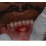 |
Figure 1 |
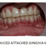 |
Figure1 b: Reduced Attached Gingiva Width |
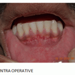 |
Figure1 c: Intra Operative |
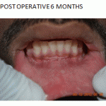 |
Figure2 a: Post Operative 6 Months |
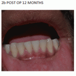 |
Figure2 b: Post Op 12 Months |
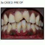 |
Figure3 a: Case 2 Pre Op |
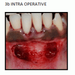 |
Figure3 b: Intra Operative |
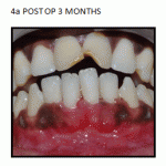 |
Figure4 a: Post Op 3 Months |
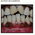 |
Figure4 b: Post Op 6 M0nths |
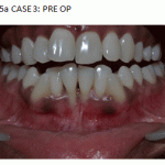 |
Figure5 a: Case 3: Pre Op |
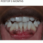 |
Figure6 a: Post Op 3 Months |
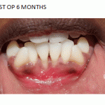 |
Figure6 b: Post Op 6 Months |
Conclusion
Mucogingival stress could be a primary factor leading to inflammation and loss of attachment in the lower anterior region. A simplistic single step approach to eliminate muscle pull causing recession and reduced attached gingival width could be more predictable in long term maintenance of health.
References
- Ainamo J, Talari A. The increase with age of the width of attached gingiva.JPeriodontal Res1976; 11(4):182-188.
- Lozdan J, Squier CA. The histology of the mucogingival junction. J Periodontal Res 1969;4(2):83-93.
- Lang NP, Loe H. The relationship between thewidth of keratinized gingiva and gingival health.JPeriodontol 1972;43(10):623-627.
- Wennstrom J L. Mucogingivaltherapy.AnnPeriodontol 1996;1 :671-701
- Friedman N. Mucogingival surgery. Tex Dent J I957; 75: 358-362.
- Miller PD Jr. A classification of marginal tissue recession.Int J Periodontics Restorative Dent1985; 5(2):8-13.
- Glickman I, Carranza FA. Clinical periodontology. 5th ed.Philadelphia: W.B. Saunders; 1979. p. 100-101.
- Beck JD. Periodontal implications: older people. Ann Peridontol1996; 1:322-357.
- Albandar JM. Global risk factors and risk indicators for periodontal diseases. Periodontology 2000 2002; 29:177-206.
- Tillis TSI, Keating JG. Understanding and managing dentine hypersensitivity.J Dent Hyg2002; 76:296-309.
- Drisko CH. Dentine hypersensitivity-dental hygiene andperiodontal considerations. Int Dent J 2002; 52supp1:385-393.
- Takei H, Azzi R, Han T. Periodontal plastic and esthetic surgery. In: Carranza F.A, editor.Clinical Periodontology. 10th ed. St. Louis: Elsevier; 2009. pp. 1005–30
- Harinder Gupta.Vestibular Extension by Edlan-Mejchar Technique Followed By Permanent Fibre Splinting. Indian Journal of Dental Sciences; March 2010 2(2): 16-19
- Lindauer SJ, Laskin DM, Taylor RS, Cushing BJ, Best AM. Orthodontists’ and surgeons’ opinions on the role of third molars as a cause of dental crowding. Am J Orthod Dentofacial Orthop 2007; 132:43-8.

This work is licensed under a Creative Commons Attribution 4.0 International License.


