How to Cite | Publication History | PlumX Article Matrix
Islam H Al-Qudah1, Ahmed Al- Radaideh2, Nader Ahmed3 and Asok Mathew 4
1General Practioner, Jordan.
2College of requirement Ajman University of science and technology, Al Fujairah.
3College of pharmacy Ajman University of science and technology, Al Fujairah.
4College of dentistry , Ajman University of science and technology, Al Fujairah.
DOI : http://dx.doi.org/10.13005/bbra/2423
ABSTRACT: With advancing diagnostic capabilities and emerging technologies it is more vivid and clear that humans are under the threat of the microorganisms. Scientific fraternity is thinking deeply now how the bacterial, viral or fungal load can be addressed.It is believed that oral bacterial challenge can pose threat to multiple organ systems. The proof is highly established and double way bacterial challenge is established in the control and etiologies of diseases like low birth weight babies, coronary diseases , diabetes mellitus, Alzheimer’s and so on. This lab study is undertaken in the microbiological lab using the specific media mitis salivarius. The study is done to the antibacterial efficacy of miswak extract in comparison with chlorhexidine, saline and distilled water. The method employed here is disc diffusion method for antibiotic sensitivity. It is noted that both chlorhexidine and miswak extract exhibited significant antibacterial property compared with saline and distilled sterile water.It is also observed that miswak extract showed better inhibition against streptococcus mutans compared with sample grown with plaque mixed population of bacterial population. The miswak extract is having comparable antibacterial efficacy with chlorhexidine. The miswak extract showed a better inhibition sensitive zone of 12mm or more when a pure colony of streptococcus mutans was applied in the mitis agar plate.
KEYWORDS: Chlorhexidine Mitis salivarius agar; Miswak; Streptococcus mutans;
Download this article as:| Copy the following to cite this article: Al-Qudah I. S, Al-Radaideh2 A, Ahmed N, Mathew A. Comparison of Antibacterial Efficacy of Miswak Extract to Conventional Mouth Washes – A Microbiological Study. Biosci Biotechnol Res Asia 2017;14(1). |
| Copy the following to cite this URL: Al-Qudah I. S, Al-Radaideh2 A, Ahmed N, Mathew A. Comparison of Antibacterial Efficacy of Miswak Extract to Conventional Mouth Washes – A Microbiological Study. Biosci Biotechnol Res Asia 2017;14(1). Available from: https://www.biotech-asia.org/?p=21345 |
Introduction
The insult of oral mucosa due to thriving oral microbes has been a challenge for oral diagnosticians and periodontologists. The greater gingival sulcular surface area exposed to these microbes is of greater importance as the bacteremia and the absorption of toxic substances as result of degradation of the cell walls of bacteria will result in a compromised general health.
This will either cause direct impact on many diseases like diabetes, coronary diseases or will result in difficulty in managing these chronic diseases. The study is done to see the possibility of developing a natural herbal based mouth wash which can give sustained reduction of harmful bacteria without affecting beneficial bacteria. Miswak is found to reduce the bacterial count of strepto coccus sp: without much effect on candia sp.1
Meswak “a chewing stick”( Salvadora persica) is a herb used to clean the gums and teeth and is usually 15 cm long and 1 cm in diameter is taken from the roots, can be further cut into suitable lengths by the user. A length of 20 cm for adults and 15 cm for children is recommended for convenience of the grip and ease of manipulation in a confined space. 2 The diameter of 1 cm, gives the stick enough strength to transmit the pressure of the cleansing action to the teeth without breaking off. The thicker sticks tend to be older and difficult to chew. The root should be whitish-brown in color; a dark brown color indicates that the Miswak is no longer fresh. A very dry Miswak can be expected to damage the gums and other oral tissues. If a stick is dry, the end for chewing should initially be soaked in tap water for 24 hours.
It should be noted that soaking for unduly long periods causes loss of active constituents and diminishes the therapeutic properties, although the mechanical effects on the teeth can still be expected to occur. These fibers should be clipped of every 24 hours.3,2,4 The World Health Organization (WHO) recommended the use of the miswak in 1986 and in 2000 an international consensus report on oral hygiene concluded that further research was needed to document the effect of the miswak.5
Streptococcus mutans is facultative anaerobic, Gram-positive coccus – shaped bacterium commonly found in the human oral cavity and is a significant contributor to tooth decay,6 the microbe was first described by J Kilian Clarke in 1924 .Twenty-five species of oral streptococci live in the oral cavity. Each species has developed specific specialized properties for colonizing different oral sites and constantly changing conditions to fight competing bacteria and to withstand external challenges. Imbalances in the microbial biota can initiate oral diseases.
Under special conditions, commensal streptococci can switch to opportunistic pathogens, initiating disease and damaging the host. Oral streptococci has both harmless and harmful bacteria. S. mutans, the microbial species most strongly associated with carious lesions, is naturally present in the human oral microbiota.
Biofilm is an aggregate of microorganisms in which cells adhere to each other or a surface. Bacteria in the biofilm community can actually generate various toxic compounds that interfere with the growth of other competing bacteria. However, there have been very few studies on how S. mutans can tolerate such exposure to various toxic substances during its growth in the oral biofilm and thus, poorly understood.6
Mitis salviarius agar is used to differentiate among species of Streptococcus and the very close genus Enterococcus (formerly in the Streptococcus genus) that are flora in the mouth. These organisms are very common in the mouth, and have been associated with dental caries, as well as agents of endocarditis.7 The sugars in this medium are sucrose and glucose, as well as the dyes trypan blue and crystal violet. Trypan blue is absorbed by the bacterial coolonies, causing them to become blue. The crystal violet, along with the 1% tellurite added to this medium, inhibit gram – bacteria as well as many other gram + bacteria. 7 Streptococcus mutans was identified by its characteristic colony morphology. The colonies were raised, convex, opaque with rough margins and granular frosted appearance and glistening bubble often accumulated on the top of the colony. After 48 hours the plates were removed from the incubator, the most countable plates were selected, i.e. having 30-200 CFU/ml.
Punit V.P has shown that there was a significant reduction in plaque score and improvement in gingival health after the use of miswak as an adjunct to tooth brushing method. The miswak stick when not used as adjunct to tooth brush was not effective as the conventional toothbrush in reducing plaque until 4th week. Later at the 6th and 8th week, there was significant difference in plaque score in toothbrush user compared to miswak user. This initial reduction in the plaque score among the miswak users could be due to Hawthorne effect.8 The Hawthorne effect is a form of reactivity, whereby subjects improve or modify an aspect of their behavior being experimentally measured simply in response to the fact that they are being studied, but not in response to any particular experimental manipulation
Aims and objectives
The aim of the study is to determine the anti-bacterial effect of miswak (Salvadora persica) extract and compared with (saline, chlorhexidine mouth wash and distilled water) on cariogenic bacteria like streptococcus mutans
Materials and Methods
The study was done in the microbiological lab of college of pharmacy, Ajman University of science and technology, Al Fujairah. The samples were collected from 30 patients of student clinics of Ajman University of science and technology, Al Fujairah with an age range of 20-45 years. The biofilm was scrapped from the labial surface of upper anterior teeth using sterile cotton and immediately transferred to the lab facility dipping in sterile distilled water for diffusion. The solution obtained after 30 minutes of dipping the plaque in the sterile vials is being used for further spreading on the mitis salivarius media. Biofilm bacteria showed growth in the first application as mixed colonies of many streptococcus bacteria. The samples were collected from the colonies of streptococcus mutans to be used in the 2nd set of mitis salivarius plate to get pure streptococcus mutans growth. Study was conducted using miswak extract, chlorhexidine , saline and distilled water on Streptococcus mutans and a general plaque sample.
Isolation of Mutans Streptococci Bacteria
One hundred microliter of undiluted samples was spread on the surface of MS-agar plates using sterile swabs. Cultures were incubated anaerobically for 48 hrs at 37°C and aerobically overnight at 37°C. Count of more than 250 colonies (104 cells/ml) was considered as positive samples.
Antibiotic Sensitivity Test
Disk diffusion method for antibiotic sensitivity test was determined following the method described by Baron et al.9 Disk paper (using disk diffusion test technique) was soaked in each of the 4 solutions for 30 minutes . Sterile forceps is used to distribute the soaked disk paper on the agar, kept in incubator for 48 hours, at 37°C in an inverted position. After this period of incubation, the diameters of inhibition zones were noted and measured by a ruler in (mm) and results were determined according to the National Committee for Laboratory Standards.10
Preparation of miswak extract
For this study, two types of miswak extract was prepared and each type was prepared with 40gm of fresh miswak which grounded into powder fig 1
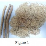 |
Figure 1 |
It is prepared by adding the miswak powder to 100ml of 50% of ethanol alcohol, and soaked for different duration, and based on that it divided into two types to see whether the soaking for long will increase or decrease the antibacterial efficacy of the solution prepared.
Type 1- Kept for 1 day.
Type 2- Kept for 10 days
By using disc diffusion test, 5 disc papers were soaked in each of the solutions (saline, chlorhexidine, distilled water and the miswak extract) for 30 minutes. Fig 2
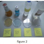 |
Figure 2 |
Then petri dishes with mitis salivarius agars are used, one for spreading of the isolated streptococcus mutans , and the other for the general plaque sample . Spreading done by used sterilized spreading loop. Fig 3
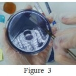 |
Figure 3 |
Both the mitis salivarius agars were then divided into 4 quadrants. A single soaked disc paper was distributed on the agars in its specific quadrant. Then the agars placed in the incubators for 24-48 hour. Fig 4
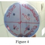 |
Figure 4 |
Each group samples were checked after the mentioned periods, to check the bacterial inhibition zone around each soaked disc paper, to evaluate the antibacterial effect of each solution on the surrounded bacteria on the agar.
The number of colonies per milliliter (CFU/ml) was determined by the following equation: Number of colonies/ml (CFU/ml) = number of colonies counted × Inverse of dilution × inverse the cultured volume (ml).11
Results
According to the anti-bacterial sensitivity test measurements, which indicate 3 main zones
Resistant Zone 1-4mm
Intermediate Zone 4-12mm
Sensitive Zone 12mm Or More
The diameter was measured for each inhibition zone that resulted on each solution on both samples for the 3 –groups. It had shown that the chlorhexidine had the best antimicrobial effect on both pure strep. Mutans sample and plaque sample. On the other hand miswak extract both type 1 & 2 had similar antibacterial effect on both the samples but the effect was more on the strep. Mutans sample. Table 1
| Group | Streptococcus bacteria | Plaque bacteria |
| Miswak ( 1 day – Type 1 ) | 14mm | 7mm |
| Chlorohixidine o.12% | 18mm | 15mm |
| Saline | 2mm | Zero |
| Distilled water | Zero | Zero |
| Miswak ( type 2 – 10 days ) | 14mm | 12mm |
| Chlorohixidine o.12% | 16mm | 17mm |
| Saline | 4mm | Zero |
| Distilled water | Zero | Zero |
Miswak extract (1 day) type 1
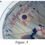 |
Figure 5 |
Show the antibacterial effect of miswak type 1 solution on the selective Strep. mutans sample fig 5
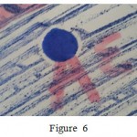 |
Figure 6 |
The antibacterial inhibition zone of miswak type 1 on the general plaque sample fig 6
Miswak extract (10 days) Type 2
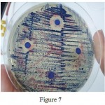 |
Figure 7 |
The fig 7 shows the effect of miswak type 2 on the selective Strep . Mutans sample .
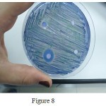 |
Figure 8 |
The fig 8 shows the effect of miswak Type 2 on the general plaque sample
Discussion
The method of cleaning with miswak is linked with religious beliefs in the Middle East, Saudi Arabia, United Arab Emirates and even Africa. The use of this herb for effective oral hygiene is recommended by WHO for prevention of oral diseases. This study is done to establish the efficacy of miswak in attaining the antibacterial activity against mixed streptococcus sp: as well as selective activity against streptococcus mutans. The study is relevant as the feasibility of development of natural mouth wash based on miswak extract is explored here with very promising results.
Darmani H et al. examined the effects of aqueous extracts of miswak on the growth of the various cariogenic microorganisms including Streptococcus mutans. The result showed inhibition in growth of Streptococcus mutans which is similar to our result.12 It was found by (Padma K Bhat ) that miswak extract had very significant detrimental effect on both the dental caries causing micro-organisms at the tested conditions. It had shown significant reduction of microbial count as compared to sterile water and saline in the present study.13 A cross- sectional study in Ghana among adults revealed higher plaque and gingival bleeding in chewing stick users as compared with toothbrush users. Another retrospective study showed that Miswak users had deeper pockets and more prevalence of periodontal diseases.14 In contrast, no differences in plaque and gingival bleeding were found between toothbrush and chewing stick users among 7-15 years old children in Tanzania. It is reported that patients using Miswak regularly show decreased gingival bleeding on probing compared with non-Miswak users.Thus, poor oral hygiene with those using chewing sticks may be a reflection of poor techniques. On the other hand, controlled longitudinal studies were more consistent.4
Al-Bayaty FH et al, 15 Shingare P et al 16 and Masoumeh K et al17 had also found the miswak extract as an effective antimicrobial agent which is comparable to our study results. Al-Bagieh reported that Benzyl iso thiocyanate inhibits the growth and acid production of streptococcus mutans. Others found that chewing sticks were effective in reducing plaque and gingival inflammation. Where properly used, miswak had been reported to be as effective as tooth brushing and was found that plaque and gingivitis were significantly reduced when miswak was used 5 times a day compared with conventional toothbrush.18 In summary, it can be concluded that miswak was as effective as a toothbrush for reducing plaque both experimentally and clinically.
Furthermore, a study by Darout et al showed that aqueous miswak extracts contained potential antimicrobial anionic components in addition to chloride and sulphate, which were thiocyanate and nitrate. They hypothesized that thiocyanate leaching out from miswak, while in the oral cavity, may lead to an elevated level of salivary thiocyanate. This, in turn, may enhance the efficacy of the salivary hydrogen peroxide–peroxidase-thiocyanate system, a known antimicrobial component of human saliva.19
The study is done with few samples and can be done with more samples to get more accurate results. The study is done to see the antibacterial efficacy of streptococcus mutans and be extended to the see the antibacterial activity against other possible pathogens.
Conclusions
The antibacterial effect is maximal in chlorhexidine solution when compared with miswak ,saline and sterile water.
The antibacterial effect is in the sensitive zone in both miswak and chlorhexidine solutions
The antibacterial inhibition zone is more in the pure streptococcus mutans samples when miswak extract is applied
The number of days the miswak extract remained after preparation had no significant effect on the disc inhibition antibiotic sensitivity test.
References
- HowaidaF. In vitro antimicrobial effects of crude miswak extracts on oral pathogens. Saudi Dental Journal. 2002;14(1):1-2.
- Ra’ed I & KhalidS A. Miswak (chewing stick); A Cultural and Scientific Heritage. Saudi Dental Journal. 1999;11( 2):2-6.
- Glass R. T. The infected Toothbrush, the infected denture, and transmission of Disease ;a review.Compendium. 1992;13(7):592–598.
- Sadhan R. I., Almas K. Miswak (chewing Stick) A Cultural and Scientific Heritage. Saudi Dental Journal. 1999;11(2):1-6.
- Wikipedia, the free encyclopedia Miswak (online) available at : http://en.wikipedia.org/wiki/Miswak#Studies accessed 05/04/2016.
- Wikipedia, the free encyclopedia, 2013.Streptococcus mutans(online) available at : http://en.wikipedia.org/wiki/Streptococcus_mutans accessed :7/3/2016.
- Reynold Microbiology, Mitis salivarius agar (online) available at: http://delrio.dcccd.edu/jreynolds/microbiology/2421/lab_manual/atlas/biol2420photoatlas_ms.htm accessed date: 10/06/2016.
- Vaibhav P. P. S and Kumar S. Clinical effect of miswak as an adjunct to tooth brushing on gingivitis .J Indian Soc Periodontol. 2012;16(1):84–88
CrossRef - Baron E. J., Finegold S. M and Peterson L. R. Genetic and physiological study on cariogenci Streptococcus species.Journal of Al-Nahrain University. 1994;11(3).1-2.
- Matthew A., et al Performance Standard for Antimicrobial Susceptibility Testing.National Committee for Clinical Laboratory Standards. 2001;11(17):41.
- Nada H. A., et al .Isolation and Identification of Mutan’s Streptococci Bacteria from Human Dental Plaque Samples.journal of al-nahrain university. 2008;11(3):2-3.
- Darmani H,,Nusayr T & Al-Hiyasat A. S. Effects of extracts of miswak and derum on proliferation of Balb/C 3T3 fibroblasts and viability of cariogenic bacteria. J Dent Hygiene. 2006;(4):62–65.
CrossRef - Kumar B. A & Ksarkar S. Assessment of Immediate Antimicrobial effect of Miswak Extract and Toothbrush on Cariogenic Bacteria ;A clinical study.Journal of advanced oral research. 2012;3(1):17-18.
- Norman S,,Mosha H. J. Relationship between habits and dental Health among rural Tanzanian children.Comm Dent Oral Epidemiol. 1989;17:317-21.
CrossRef - Al-Bayaty F. H & AI-Koubaisi A. H & Ali N & Abdulla M. A. Effect of mouth wash extracted from Salvadorapersica (Miswak) on dental plaque formation;A clinical trial. Journal of Medicinal Plants Research. 2010;4(14):1446-54.
- Shingare P & Chaugule V. Comparative evaluation of antimicrobial activity of miswak, propolis, sodium hypochlorite and saline as root canal irrigants by microbial culturing and quantification in chronically exposed primary teeth. GERMS. 2011;1(1):12-21.
CrossRef - Masoumeh K & Solmaz A & Mahdi K & Fariborz V.Comparison of the Antimicrobial Effects of Persica Mouthwash and 0.2% Chlorhexidine on AggregatibacterActinomycetemcomitans of Healthy Individuals and Patients with Chronic Periodontitis.Research Journal of Medical Sciences. 2012;6(1):18-21.
CrossRef - M. The effectiveness of chewing Stick Miswak on Plaque removal.Saudi Dental Journal. 2006;18(3):1-2
- Darout I. A & Christy A. A & Skaug N &Egeberg P. K. Identification and quantification of some potentially antimicrobial anionic components in miswak extract. Indian J Pharmacol. 2000;32:11–4.

This work is licensed under a Creative Commons Attribution 4.0 International License.


