How to Cite | Publication History | PlumX Article Matrix
Russel R. Ghanim , M. R. Mohammad and Adi M. Abdul Hussien
Branch of Applied Physics, Department of Applied Science, University of Technology, Baghdad-Iraq.
Corresponding Author E-mail: russull.rushdi@gmail.com
DOI : http://dx.doi.org/10.13005/bbra/2669
ABSTRACT: Graphene oxide (GO) nanosheets were prepared by a novel simplified Hummer's method. The morphological and cross section images of GO have been tested with field emission-scanning electron microscope (FE-SEM). The antibacterial activity of GO nansheets against Escherichia coli (E. coli) and Staphylococcus aureus (S. aureus) were investigated as a model for Gram-negative bacteria and Gram-positive bacteria respectively. Bacteriological tests were performed by agar well diffusion assay with different concentrations of GO nanosheets and the bacterial morphological change of two bacterial species has been studied by scanning electron microscope (SEM) before and after treated with GO nanosheets. These sheets have been approved to be an effective bactericide. The antibacterial activity of the nanosheets dispersion was measured by agar well diffusion method. Scanning electron microscopy (SEM) was used to investigate the biocidal action of this nanoscale material. The nanosheets of GO have shown a high antibacterial activity against Gram-positive bacteria. The results of the present work offer a novel assay to prepare GO nanosheets were it could be used as novel antibacterial agent in future for different areas of biomedical and pharmaceutical sciences, like biosensing, antibiotics, imaging, and drug delivery.
KEYWORDS: Agar Well Diffusion Assay; Antibacterial Activity; Graphene Oxide; SEM
Download this article as:| Copy the following to cite this article: Ghanim R. R, Mohammad M. R, Hussien A. M. A. Antibacterial Activity and Morphological Characterization of Synthesis Graphene Oxide Nanosheets by Simplified Hummer's Method. Biosci Biotech Res Asia 2018;15(3). |
| Copy the following to cite this URL: Ghanim R. R, Mohammad M. R, Hussien A. M. A. Antibacterial Activity and Morphological Characterization of Synthesis Graphene Oxide Nanosheets by Simplified Hummer's Method. Biosci Biotech Res Asia 2018;15(3). Available from: https://www.biotech-asia.org/?p=30850 |
Introduction
Applications of the nanotechnology to health care can be termed as “nanomedicine” which requires a combination of various disciplines including physics, chemistry, biology and medicine.1 High chemical reactivity, ability to form aggregates, diffusivity, and the high surface to volume ratio of nanoscale materials is related with a number of novel and eligible properties as compared to the corresponding bulk materials. These properties involve chemical, mechanical, electrical and optical characteristics such as light absorption and conductivity, as well as catalytic and biological activity.2-4 During the last ten years, synthesis and characterization of graphene based materials of sp2 single atom sheet of bonded carbon atoms in a closely packed honeycomb of two dimension lattice, is regarded one of the most attractive nanostructures have evoked much interest because of their variety electronic, thermal, optical, mechanical, and magnetic properties.5,6 Different carbon materials, such as graphene, graphene oxides (GO) and reduced graphene oxides (RGO) were intensively paid attention during last decade, as potential antimicrobial agents in tissue engineering biomaterials with minimal toxicity to live cells. Their biocompatibility, high mechanical strength, high surface area, also the ability to induce sustained stem cell growth and differentiation to various lineages which are regarded additional advantages.7 Carbon based nanomaterials, such as two dimensions (graphene nanosheets), one dimension (Carbon nanotubes), and zero dimension (fullerenes) are widely used in different fields of nanotechnology. Graphene nanosheets can degrade the bacteria. They also reduce cell viability, which can attack the cell membranes and extract great amounts of phospholipids due to strong dispersion interactions between graphene and lipid molecules.8 The GO has a large surface area (high surface to volume ratio), and good dispersibility in most solvents. The presence of large quantities of oxygen containing functional groups on the sheet basal plane and sheet edge of GO makes it extremely hydrophilic and provides the capability to apply GO in the biological environment. The GO nanosheets with these many reactive groups can interact strongly with cells and easily penetrate or cover cell surfaces, showing higher affinity for bacteria.9,2 The E. coli and S. aureus are the common bacterial species and the main cause of infectious diseases in humans and animals.10 Therefore, it is significant to look for new antibacterial materials as alternative of antibiotics for therapy these bacterial strains.
In the current work, the common method for preparing GO nanosheets is Hummer, which is followed since 1958 by W. S. Hummers et al.,11 This method have been simplified in this study to be more facile, require fewer apparatus (just magnetic stirrer), and hence lower cost. The antibacterial susceptibility of GO nanosheets for the Gram-negative (E. coli) and the Gram-positive (S. aureus) were investigated in the laboratory with different concentrations by using the most often performed way that is the well diffusion method. This method is well documented and standard zones of inhibition have been determined for susceptible and resistant values. The antibacterial activity also achieved by studying the influence of GO nanosheets on morphology of bacterial cell wall by using SEM.
The aims of the present study were to synthesize GO nanosheets with a novel method by simplifying the main Hummer’s method, and to test the antibacterial activity of these nanostructures.
Materials and Experimental Techniques of Graphene Oxide Nanosheets
Chemicals and Reagents
Nanostructured GO sheets have been prepared by a novel method using a simplified Hummer’s method from the laboratory Graphite powder, which has been supplied by Sigma Aldrich Company (Germany). The Sulfuric acid (H2SO4) has been supplied by Schwefelsäure Company (Netherlands) of purity 95-97%, Potassium permanganate (KMnO4) of 99% purity has been supplied by GCC Company (England), and finally, Hydrogen peroxide (H2O2) of 37% purity has been supplied by United Horizon Company (USA).
Instrumentation
The FE-SEM morphology, microstructural features, and cross section images of dried GO drops on glass substrates have been investigated by using type (Hitachi FE-SEM model S-416, Japan). An accelerating voltage of 20 kV was used for secondary electron imaging (SEI) of 1.5 μm thickness of gold (Au) coated samples.
Preparation of Graphene Oxide by Using a Simplified Hummer’s Method
The GO nanosheets have been prepared, depending on Hummer’s method, which is an easy and efficient technique since 1958. This method has been simplified to prepare the GO with a novel method without a need to add sodium nitrate (NaNO3), and at room temperature without a need to water bath at temperatures of 35°C and 98°C. GO nanosheets have been prepared by chemical exfoliation of 1 g of graphite powder (212 μm mesh) has been added to 100 ml of high concentrated H2SO4 which acts as strong oxidant medium under magnetic starrier. In order to prevent over heat and explosion, the KMnO4 (3 g) has been gradually added to the solution placed in ice bath to keep temperature under 20°C. To obtain fully oxidization from graphite to graphite oxide, the above reaction has been continued for about 3 days. Drops of H2O2 (5% v/v) has been added to the solution, in order to destroy the excess of KMnO4, and hence to terminate the oxidation process. For purification, the mixture was washed by rinsing and centrifugation [Gemmy Industrial Corporation (Taiwan)] with speed 8000 rpm several times with 1 M of HCl solution to remove the metal ions. Then, it was washed repeatedly with deionized water to eliminate the acid in the mixture. Ultrasonic apparatus has been used, for 20 min to convert graphite oxide to GO,12 model (1740QT) supplied by VGT Company (China). A vacuum oven at 80ºC has been used to dry the sample by using (VO-27) oven supplied by Hysc Company (Korea), in order to obtain GO as a powder.
Antibacterial Activity Assay
Microorganisms and Required Materials
An E. coli isolate has been supplied by the (Medical Microbiology Laboratory, Branch of Biotechnology, Department of Applied Science, University of Technology, Baghdad – Iraq) and S. aureus has been provided by Nanotechnology Center (Microbiological Laboratory, University of Technology, Baghdad – Iraq).
The used Materials for antibacterial activity of GO nanosheets were nutrient broth (Himedia, India), Mueller–Hinton agar (Lab M 008, UK), petriplates, and cotton swabs.
Preparation of Inoculums
Nutrient broth (1.3 g) in 100 ml of distilled water (D.W.) has been prepared in two universal tubes and sterilized. In one universal tube clinically isolated strain of E. coli, has been inoculated and in the other universal tube clinically isolated strain of S. aureus has been added. The bacterial cultures inoculated nutrient broth has been kept on incubated for 24 h at 37°C.
Experimental Techniques
Agar well diffusion assay, and SEM technique were used for antibacterial activity of GO nanosheets.
Agar Well Diffusion Assay
The antibacterial activity of the GO nanosheets were tested against two types of bacteria: E. coli (Gram-negative bacteria), and S. aureus (Gram-positive bacteria) by using agar well diffusion method. This assay includes weighting of 3.8 g of Mueller Hinton agar and dissolved into 100 ml of D.W. The medium, which was prepared, has been put in an autoclave, and it passes through cycle of sterilization. Nutrient agar (about 20 ml) has been poured into sterilized Petri dishes and allowed to solidify. Bacterial strains were taken from stock bacterial culture by sterile swab, where inoculums of bacterial were spread on Mueller Hinton agar. Then, 6 mm diameter wells were holed into the agar with the sterile tip. The GO nanosheets have been loaded with equal volume (60 μl) with different concentrations 1000, 750, 500, and 250 μg/ml on the plates. The plates, which contain the test organisms, were incubated for 24 h at 37°C. As the bacteria have been grown to take a confluent lawn shape, the inhibition of growth could be measured as the expansion of the clear zones surrounding the holes on the Petri dish. The inhibition zone (IZ) diameters have been recorded by using a meter ruler, whereas, the average value for each well has been calculated. All of these tests have been achieved in triplicate. Distilled water has been used as a negative control.
Morphological Changes of Bacterial Cells between Untreated and Treated with Graphene Oxide Nanosheets
The SEM technique has been used to observe any morphological changes of bacterial cells. Four groups of specimens are E. coli and S. aureus cells were tested after and before treatment with GO nanosheets. Bacterial cells of E. coli and S. aureus were treated with GO nanosheets for 4 h. The prepared samples for SEM technique, the treated and untreated bacterial cells of E. coli and S. aureus with GO nanosheets, were centrifuged at 3000 rpm for 5 min and then washed 3 times with phosphate buffer saline (PBS, 50 mM, pH 7.3). Later on, a suspension of thin smear was spread on a silicon wafer slide and dried at room temperature. In order to make sure of good SEM technique performance, the samples were sputtered with Au for 5 min, to create a 2 nm thickness of Au layer. The samples were measured by using a scanning electron microscope (SEM model VEGA3LMU, TESCAN Company, USA) in the (Department of Production Engineering and Metallurgy, University of Technology, Baghdad – IRAQ) was operated at an accelerating voltage of 20 kV to observe the morphological changes of treated bacteria cells compared with that untreated.
Results and Discussion
Field Emission-Scanning Electron Microscope of Graphene Oxide Nanosheets
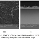
Figure (1 a) shows the morphology of GO nanosheets which represents an ultrathin and homogeneous of the GO film with a corrugated surface. However, from figure (1 b), it can be seen that the cross section photograph of GO has a layered structure with tightly packed.
 |
Figure 1: FE-SEM of the synthesized GO nanosheets. (a) The morphology image. (b) The cross section image.
|
Antibacterial Activity of Graphene Oxide Nanosheets
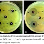
The antibacterial activity of GO nanosheets has been studied by using the agar well diffusion assay against both E. coli and S. aureus. The results of IZ against E. coli and S. aureus bacteria at different concentrations of GO nanosheets 1000, 750, 500, and 250 μg/ml have been shown in figure (2). The highest concentration showed the strongest antibacterial activity. It is clearly, that the antibacterial activity of GO nanosheets with higher concentrations had greater inhibitory effects for both Gram-negative and Gram-positive bacteria strains. The effect of nanosheets on Gram-positive bacteria was higher than Gram-negative bacteria, which are seemed to have a little resistant to the nanosheets as shown in table (1).
In general, small size nanoparticles show better antibacterial activity, because decreasing of volume will cause increasing of surface area, and hence increase the antibacterial activity. The cell membrane has nanosized pores which allow the smaller nanoparticles easily to penetrate the cell and reach to the nuclear content of bacteria (reverse osmosis).13
Table 1: Inhibition zone of GO nanosheets against E. coli and S. aureus.
| Concentration (μg/ml) | 1000 | 750 | 500 | 250 |
| Inhibition Zone of E. coli (mm) | 19 | 17 | 16 | 15 |
| Inhibition Zone of S. aureus (mm) | 25 | 20 | 19 | 18 |
 |
Figure 2: Antibacterial activity of GO nanosheets against (A) E. coli and (B) S. aureus, where C. represents control (D.W.) and 1, 2, 3, and 4 represent nanosheets with concentrations of 1000, 750, 500, and 250 μg/ml, respectively.
|
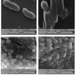
SEM technique has been used to test the changes in cell morphology after contacting to GO nanosheets. In order to explain the effect of GO nanosheets on the bacteria, GO nanosheets can damage the bacterial cell membrane due to the penetration. Hence the SEM technique have been used to measure the morphologies of E. coli and S. aureus bacteria after and before treating with the GO nanosheets, as illustrated in figure (3).
In E. coli bacteria, the SEM images of control cells were typically rod shape (figure 3 A) which represents a uniform bacteria cell surfaces. However, in the GO nanosheets treated bacterial cells (figure 3 B) it is noted that there is an indication of occurred damage, and hence structural changes on bacterial cell membrane.
In S. aureus bacteria, the SEM image of control cells has a typically grape shape (clusters) (figure 3 C), the cell surface was intact, and each cell size was almost same. However, after GO nanosheets treatment (Figure 3 D), an irregular shape of the bacterial cell was appeared, which indicates a structural changes and damage on bacterial cell surface. An increasing of penetration of cell membrane and/or leakage of the cell contents which could be caused by reactive oxygen species (ROS).14
 |
Figure 3: SEM micrograph of E. coli (A, B) and S. aureus (C, D). (A, C: control; B, D: treated with GO nanosheets).
|
The images of figure (3) were revealed the morphological variation of the two kinds of bacteria membranes and the damage of the cells structure.
The observed differences are due to many reasons such as the primary variance between the Gram-negative and Gram-positive bacteria with respect to the nature of their cell wall, as well as, the Gram-negative bacteria have an additional outer membrane comprising of lipopolysaccharide which protects the peptidoglycan layer from chemical attacks.1
Structural variations in the bacterial cell wall led to the classification of bacteria into Gram-negative and Gram-positive. Where, the last bacterial cell wall involves a thick peptidoglycan layer and another coat of polysaccharide usually formed from teichoics and teichuronic acids. However, the cell wall of Gram-negative bacteria has more complexity, include thin layers of peptidoglycans, lipoproteins, lipopolysaccharides and an outer membrane. This structure explains, why Gram-positive would be more influenced by nanoparticles than Gram-negative bacteria.15
It has been suggested that the higher capability of Gram – positive bacteria could be associated to the differences of cell wall structure, cell physiology, metabolism or degree of contact.
An E. coli bacterium has resistant to the antibacterial influence of the GO nanosheets as compared to a S. aureus bacterium. This can be explained by two reasons. Firstly, the existence of a number of small channels of porins within the outer membrane of E. coli may help block the entrance of the nanoparticles into the bacterial cell, making them more complicated to inhibit than S. aureus. Secondly, the smaller dimension of S. aureus (sphere, about 0.5-1 μm) may partly account for the more intimate contact with the nanoparticles, making the antibacterial activity of the S. aureus more efficient than that for the E. coli (rod, about 0.3-1× 1-6 μm).16
Generally, the cell membrane appears to be a primary target of the cytotoxicity of GO nanosheets. Membrane damage may be caused by the atomically sharp edges of graphene, which could penetrate the cell membrane and physically disrupt its integrity. The oxidative nature of GO also presents as another cause of membrane damage for bacterial cells that exposed to GO.17,18
Conclusions
In this study, the GO nanosheets have been prepared by the simplified Hummer’s method; it is facile and inexpensive chemical way. The morphology and cross section images of FE-SEM measurements of GO nanomaterials confirmed the existence of layered structure. These synthesis GO nanosheets have been tested as antibacterial activity for Gram-negative and Gram-positive bacteria. The GO nanomaterial shows antibacterial properties, and the obtained results allowed to formulate the conclusion that nano-GO may inhibit bacterial growth. The IZ test has been found that the antibacterial activity of the GO nanosheets is increased with increasing nanosheet concentration in well diffusion method, where the greatest inhibitory influence at 1000 μg/ml for two types of bacteria and were more efficient against Gram-positive than Gram-negative bacteria. The surface morphological changes have been examined by using SEM technique; the treated bacterial cells were considerably changed and showed great damage compared to that untreated (controls) for two species. Overall, the optimal antibacterial activity of GO nanosheets was observed against S. aureus bacteria, in comparison with E. coli bacteria. The results of the current study confirmed the GO nanosheets have been prepared successfully by novel way and can be used as antibacterial material for biomedical and pharmaceutical applications.
Acknowledgments
The authors are thankful to the Department of Applied Science, University of Technology, Baghdad – IRAQ for supply the excellent facilities. Also we express our thanks to Assist. Prof. Dr. Majid S. Jabir for his useful help.
References
- Krishnamoorthy N., Umasuthan R., Mohan J. L and Kim S. J. Investigation of the Antibacterial Activity of Graphene Oxide Nanosheets. Science of Advanced Materials. Korea. 2012;4:1-7.
- Veerapandian L., Zhang K. K and Yun K. Surface Activation of Graphene Oxide Nano sheets by Ultraviolet Irradiation for Highly Efficient Anti-bacterials. Nanotechnology journal. 2013;24:1-11.
- Doskocz K., Affek and Radziwiłł M. Z. Effects of Aluminium Oxide Nano particles on Bacterial Growth. E3S Web of Conferences. 2017;17:1-7.
CrossRef - Alananbeh M., Al-Qudah Z., El-Adly A., Refaee A .W.J. Impact of Silver Nano particles on Bacteria Isolated from Raw and Treated Wastewater in Madinah, KSA. Arab J SciEng. 2017;42:85–93.
CrossRef - Sharma M., Varshney S. S., Nanda H. J., Shin N. K., Yi D. K., Chae K. H and Won S. O. Structural Electronic Structure and Antibacterial Properties of Graphene Oxide Nano-sheets. Chemical Physics Letters. 2018;698:85–92.
CrossRef - Akhavan and Ghaderi E. Toxicity of Graphene and Graphene Oxide Nanowalls against Bacteria. ACS Nano journal. 2010;4(10):5731–5736.
CrossRef - Vladkova G.,Ivanova I. A., Staneva A. D., Albu M. G., Shalaby A. S. A., Topousova T. I and Kostadinova A. S. Preparation and Biological Activity of New Collagen Composites Part II: Collagen/Reduced Graphene Oxide Composites. J Arch Mil Med. 2017;5(1):1-9.
CrossRef - Vass Z., Deák Z., Paul K., Kovács S Vass I. Interaction of Nano particles with Biological Systems. Interaction of nano particles. 2015;59(2):225-245.
- Gao J., Wu X., Rena X., Tana T., Hayatd A., Alsaedid C., Chenga and Chena C. Impact of Graphene Oxide on Antibacterial Activity of Antibiotics against Bacteria. Environ. Sci. Nano. 2017;4(5):1016-1024.
CrossRef - Bannerman D. D., Paape M. J., Lee J. W., Zhao X., Hope J. C and Rainard P. Escherichia coli and Staphylococcus aureus Elicit Differential Innate Immune Responses Following Intramammary Infection. Clin Diagn Lab Immunol. 2004;11(3):463-472.
CrossRef - Hummers S and Offeman R. E. Preparation of Graphitic Oxide. J. Am. Chem. Soc. 1958;80(6):1339-1339.
CrossRef - Li X. Z. , Ren T and Heide E. v. d. he Preparation of Graphene Oxide and Its Derivatives and Their Application in Bio-Tribological Systems. Lubricants. 2014;2:137-161.
CrossRef - Paul D., Mittal R. Nature of Interaction between Metal Nano particles (Ag) & Bacterial Cell (E.Coli), a dissertation Submitted in Partial Fulfillment of the Requirements for the Degree in Bachelor of Technology in Biotechnology Engineering. National Institute of Technology Rourkela. 2012.
- Kim S., Hwan H. S., Lee D. S., Ryu S., Choi J and Lee D. S. Antibacterial Activity of Silver – nanoparticles against Staphylococcus aureus and Escherichia coli. Korean J. Microbiol. Biotechnol. 2011;39(1):77–85.
- Neto M., de Almeida K. C., Macedo M. L. R., Franco O. L. Understanding Bacterial Resistance to Antimicrobial Peptides: From the Surface to Deep Inside. Biochimica et Biophysica Acta. 2015;1848:3078–3088.
CrossRef - Li Q., Chen J., Zhao & Urmila K. Enhancing the Antimicrobial Activity of Natural Extraction Using the Synthetic Ultrasmall Metal Nano particles. Sci Rep. 2015;5.
- Franc-ois A., de Faria F., Nejati S and Elimelech M. Antimicrobial Properties of Graphene Oxide Nanosheets: Why Size Matters. ACS NANO. 2015;9(7):7226–7236.
CrossRef - Nanda S. S., Yi D. K and Kim K. Study of antibacterial mechanism of graphene oxide using Raman Spectroscopy. Scientific Reports. 2016;6:1-12.
CrossRef

This work is licensed under a Creative Commons Attribution 4.0 International License.


