

How to Cite | Publication History | PlumX Article Matrix
Fungal Profile of Sugar Cane (Saccharum Officinarum L.) Pathogens Produced in Côte D’ivoire
Djédji Okoue1 , Fulgence Yao Koffi2*, Armist Amoa Tehua3, Ibrahima Kebe1 and Mireille Waze Aimée Alloue-Boraud1
1Department of Food Science and Technology, Nangui Abrogoua University, Abidjan, Côte d’Ivoire.
2Department of Biochemistry-Genetics, Peleforo GON COULIBALY University, Korhogo, Côte d’Ivoire.
3Department of Plant Biology, Peleforo GON COULIBALY University, Korhogo, Côte d’Ivoire.
Corresponding Author E-mail:fulgencekoffi85@yahoo.fr
ABSTRACT: Sugar cane occupies an important place in the Ivorian economy. However, it is subject to fungal attacks during its production, leading to enormous economic losses for farmers and manufacturers. The aim of this work was establish profile of pathogenic fungal strains of sugar cane in Côte d’Ivoire. To do this, 250 sugarcane samples composed of leaves, stems and shoots of sugarcane presenting pathologies were collected, then phenotypic and molecular identifications of the pathogenic fungal strains were carried out by PCR followed by sequencing. After identification, a pathogenicity test was carried out to demonstrate the involvement of these fungal strains in sugar cane pathologies. Three (3) fungal species including Fusarium verticillioides, Fusarium subglutinans and Schizophyllum commune were identified. Fusarium verticillioides is believed to be the agent responsible for red movre disease and Fusarium subglutinans responsible for “Pokkah Boeng” disease. The fungus Schizophyllum commune is thought to be responsible for anthrax.
KEYWORDS: Fusarium subglutinans; Fusarium verticillioides; Pathogen; PCR; Sugarcane; Schizophyllum commune
| Copy the following to cite this article: Okoue D, Koffi F. Y, Tehua A. A, Kebe I, Boraud M. W. A. A. Fungal Profile of Sugar Cane (Saccharum Officinarum L.) Pathogens Produced in Côte D’ivoire. Biotech Res Asia 2025;22(1). |
| Copy the following to cite this URL: Okoue D, Koffi F. Y, Tehua A. A, Kebe I, Boraud M. W. A. A. Fungal Profile of Sugar Cane (Saccharum Officinarum L.) Pathogens Produced in Côte D’ivoire. Biotech Res Asia 2025;22(1). Available from: https://bit.ly/3ReJjAS |
Introduction
Ivorian sugar production amounts to 330,000 t. The cultivation of sugar cane is subject to several constraints. The pathologies associated with this crop are caused by viruses, stem-boring insects, bacteria and fungi1. The monoculture practiced for several years in the sugar complexes leads to a decline in soil fertility. All of these factors contribute to a drop in production2. Biotic constraints have caused reductions in yield in the sugar industries of Côte d’Ivoire, notably those of Borotou-Koro and Zuénoula. Fight against these pathogens, pesticides, especially synthetic fungicides such as benzimidazoles, triazoles and strobilurins, are mainly used. However, the excessive use of these products in sugar cane plantations could encourage the appearance and proliferation of resistant strains, leading to an increase in diseases3. Faced with these harmful effects, it is essential to precisely identify and know the pathogenic fungal profile, in order to effectively fight against these phytopathogens responsible for serious pathologies. The fungal agents responsible for these diseases are often unknown. It is within this framework that this study falls, the general objective of which is to establish the profile of pathogenic fungal strains of sugar cane in Côte d’Ivoire.
Materials and methods
Material
Organs (stems, leaves and shoots) of sugar cane presenting pathologies were collected in the village plantations of Borotou-koro (08°31 North latitude and 7°17 West longitude) and Zuénoula (7°30 and 7°40 North latitude, and between 6°50 and 6°15 West longitude) in Côte d’Ivoire.
Sampling
Samples of shoots, leaves and stems showing symptoms of pathology were collected. Thus, 5 samples consisting of 5 leaves, 5 stems and 5 shoots presenting pathologies were collected per plantation. Or 50 samples at a rate of 25 samples per production area. The collected samples were packaged in sterile stomacher bags, labeled, sealed then kept in a cooler containing cold accumulators where the temperature was from 4°C.
Phenotypic identification of pathogens
Isolation was carried out according to the method of Silué et al4. Thus, the sugarcane organ fragments were sterilized in ethanol (70%) during one minute, immersed in 3% bleach for 3 minutes, and rinsed with sterile distilled water three times. The fragments collected were inoculated by direct contact on medium Potato Dextrose Agar with Chloramphenicol. The culture were then incubated at 27±1°C for 24 to 72 hours. Macroscopic identification was carried out according to the method of Botton et al5 by examining the culture. The cultural characteristics determined were the texture, the color of the thallus and the color of the underside of the crop. As for the microscopic identification, it was carried out according to the method described by Guiraud6 taking a filament using sterile tweezers then placed in a drop of methylene blue, placed on object slide and covered with a coverslip then observed under a LEICA DM750 optical microscope with an X40 objective. The characteristics observed were the appearance of the mycelium (compartmentalized or not), the presence and shape of the spores (oval, spherical, round, etc.), the shape of the conidial heads and the size of the conidiospores (short or long).
Molecular identification of pathogens
DNA extraction
Fungi colonies were collected and bubbled in 1 ml of sterile water. The suspension thus obtained is centrifuged at 12,000 rpm for one minute. The pellet of this suspension was collected and suspended in 200 μl of Chelex 100 (Bio-Rad) previously prepared following to the manufacturer’s instructions. The new suspension obtained was homogenized using a vortex for ten seconds. Heating in a water bath at 100°C for eight minutes was carried out followed by sudden cooling in ice. This thermal shock allows the lysis of cells and the release of DNA. A new homogenization step using a vortex for ten seconds was carried out at 12,000 rpm for two minutes. In the base are the proteins captured by the Chelex 100 and in the supernatant is the DNA. DNA has been preserved at -20°C for PCR amplification.
Polymerase Chain Reaction
Primers ITS1 (5’-TCCGTAGGTGAACCTGCGG-3’) and ITS4 (5’ TCCTCCGCTTATTGATATGC-3’) were used. For the preparation of the reaction mixture, an Invitrogen kit was used. This kit consists of Taq DNA polymerase, polymerase buffer and MgCl27. dNTPs and primers were also used. Final volume of amplification reaction was 50 μl. PCR medium was composed of 26.35µL of MilliQ Water, 5µL of ITS1, 2.5 μL of ITS4, 0.25 μL of Taq polymerase (Promega) and 10 μL of template DNA. The amplification reaction was made in a thermal cycler (Techne® Prime Thermal Cycler Range, USA) according to the program described by Bessadat et al8. The first step is that of initial denaturation. It is done at 94°C for five minutes. The second stage contains 35 cycles, each of which is composed of a denaturation phase at 94°C for thirty seconds, annealing phase at 55°C for thirty seconds and elongation phase at 72°C. Finally, the third stage is that of the final elongation which is done at 72°C for ten minutes.
Profile visualisation
The PCR profile were revealed by electrophoresis on 1.5% agarose gel. For gel preparation, the agarose was suspended in 50 ml of 0.5X TBE. The suspension was heated and left to room temperature for five minutes then a drop of ethidium bromide was incorporated before pouring it into a gel holder. Once solidified, the gel was immersed in an electrophoresis tank containing 0.5X TBE. 10 μl of amplified fragments were mixed with 2 μl of loading buffer (50% TE, pH 8; 50% glycerol; bromophenol blue) before being loaded into the gel wells. Profile was visualized using a transilluminator (Vilber Lourmat) under UV illumination after migration at 90 Volts for 90 minutes. The size of the DNA fragments was determined using the Reddy Run Super ladderlow 100 bp molecular size marker (Thermo Scientific) used as a reference.
Sequencing
Sequencing was carried out from the rDNA products amplified with an automated sequencer. The primers used for the amplification of the regions studied were also used as sequencing primers. All DNA sequences obtained during sequencing were combined in a database (GenBank), used for the reconstruction of taxonomic relationships between the species studied. The homology of related sequences was investigated using the bioinformatics search tool BLAST. Multiple sequence alignment was carried out using Clustral W software9.
Pathogenicity of isolated fungi
Preparation of fungal inoculum
Ten milliliters (10 mL) of sterilized distilled water were added to 14-day-old mycelial cultures of pathogenic fungi. The surface of these cultures was scraped using a sterilized metal spatula. The resulting solution was homogenized in a test tube using a vortex. A few drops (200 µL) of this suspension were used to fill the wells of a Malassez cell in order to estimate the number of spores. This number was adjusted to 106 spores/ml by dilution or by increasing the spore concentration of the solution by returning the spore solution to another mycelial culture. One milliliter (1 mL) of a 1% glucose and agar solution is added to the spore suspension before inoculation. The role of this solution is to facilitate the adhesion and germination of spores on the leaves10.
Infection of sugarcane seedlings with spore suspensions of pathogenic fungi
Using a syringe, the underside of the leaves of sugarcane seedlings was infected with 1 mL of inoculum of the pathogenic fungus. The greenhouse was regularly humidified (twice a day) by watering using a watering can in order to maintain the high humidity level (95 – 100%). The purpose of humidification was to facilitate the germination of spores of the fungal pathogen. The evolution of fungal disease symptoms was monitored regularly for three (3) months. Symptomatic organ samples were collected for the isolation of inoculated pathogenic fungi10.
Results
Microscopic pathogenic fungi of sugar cane
Sugar cane samples are attacked by molds presenting colonies of various appearances, textures and colors. Thus, 129 fungal isolates were isolated taking into account the resemblance of thalli and spores. Phenotypic identification according to the macroscopic characteristics of the colonies and on the basis of the microscopic characteristics of the mycelium and conidia or spores made it possible to highlight 2 genera, namely Fusarium and Schizophyllum (Table 1).
Table 1: Phenotypic characteristics of isolated genera.
| Genera | Characteristics |
| Schizophyllum | Rapid growth, a cottony appearance, abundant mycelial texture of white color on the upper side and yellow on the back of the culture box. Septate mycelium and spherical unicellular formations or teliospores. |
| Fusarium 1 | A cottony appearance, a white mycelial texture on the surface and on the back of the box with a flaky appearance. Mycelium is septate with the presence of lunar crescent-shaped conidia. |
| Fusarium 2 | White color on the surface and produces abundant false aerial buds and the back of the box has a flaky appearance. Mycelium is septate with the presence of conidia. |
| Fusarium 3 | Flaky appearance. The mycelium is white in color with the presence of orange-colored spores. During growth, the orange color diffuses across the entire back of the box. The hyphae are septate and branched with the presence of ellipsoidal hyaline conidia |
Molecular characteristics of sugarcane pathogenic fungal isolates
Sequence analysis made it possible to identify 3 species with similarity rates of 99 % (Table 2). Thus, we obtained Schizophyllum commune (I), Fusarium verticillioides (II) and Fusarium subglutinans (III).
Table 2: Fungi strains identified from sugar cane samples
| Groups | Nc | %H | Corresponding species | Sequences |
| i | 606 | 99.49% | Schizophyllum commune | GGAAGGATCATTAA CGAATCAAACAAGT TCATCTTGTTCT GATCCTGTGCAC CTTATGTAGTCCC AAAGCCTTCACGG GCGGCGGTTGA CTACGTCTACCTCA CACCTTAAAGTAT GTTAACGAATGT RATCATGGTCTTGA CAGACCCTAAAA AGTTAATACAACTT TCGACAACGGATC TCTTGGCTCTCGCATCGAT GAAGAACGCAGCG AAATGCGATAAGTAATGT GAATTGCAGAATTCAGT GAATCATCGAATCTTTGAACG CACCTTGCGCCCTTTGGTAT TCCGAGGGGCATGCCT GTTTGAGTGTCATTAA ATACCATCAACCCTC TTTTGACTTCGGTCTCGAG AGTGGCTTGGAAGTGG AGGTCTGCTGGAGCC TAACGGAGCCAGCTCCTC TTAAATGTATTAGCGGAT TTCCCTTGCGGGATCGC GTCTCCGATGTGATAATTT CTACGTCGTTGACCATCTCGGG GCTGACCTAGTCAGTTTCAAT AGGAGTCTGCTTCCAACCGT CTCTTGACCGAGACTAGC GACTTGTGCGCTAACTTT TGACTTGACCTCAAATCAGGT AGGACTACCCGCTGAAC TTAAGCATATC |
| ii | 513 | 100 % | Fusarium verticillioides | GGAGGGATCATTACCGAG TTTACAACTCCCAAACCCC TGTGAACATACCAATTGTT GCCTCGGCGGATCAGCCCGCTCC CGGTAAAACGGGACGGCC CGCCAGAGGACCCCCAA ACTCTGTTTCTATATGTAA CTTCTGAGTAAAACCATAA ATAAATCAAAACTTTCAACAA CGGATCTCTTGGTTCTGGCATCG ATGAAGAACGCAGCAAAAT GCGATAAGTAATGTGAATTG CAGAATTCAGTGAATCAT CGAATCTTTGAACGCACA TTGCGCCCGCCAGTATTCTG GCGGGCATGCCTGTTCG AGCGTCATTTCAACCCTCAA GCCCAGCTTGGTGTTGGG ACTCGCGAGTCAAATCGCG TTCCCCAAATTGATTGGCGG TCACGTCGAGCTTCCATA GCGTAGTAGTAAAACCC TCGTTACTGGTAATCGTCGC GGCCACGCCGTTAAACC CCAACTTCTGAATGTTGACCT CGGATCAGGTAGGAATAC CCGCTGAACTTAAGCAT |
| iii | 519 | 100 % | Fusarium subglutinans | TGCGGAGGGATCATTACCGA GTTTACAACTCCCAAACC CCTGTGAACATACCAATTG TTGCCTCGGCGGATCAGC CCGCTCCCGGTAAAACGGG ACGGCCCGCCAGAGGACCC CCAAACTCTGTTTCTATATGTAA CTTCTGAGTAAAACCATAAAT AAATCAAAACTTTCAACAACG GATCTCTTGGTTCTGGCATCG ATGAAGAACGCAGCAAAATGC GATAAGTAATGTGAATTGCAGA ATTCAGTGAATCATCGAATCTT TGAACGCACATTGCGCCCG CCAGTATTCTGGCGGGCAT GCCTGTTCGAGCGTCATTTCA ACCCTCAAGCCCAGCTTGGTGT TGGGACTCGCGAGTCAAATCGCGT TCCCCAAATTGATTGGCGGT CACGTCGAGCTTCCATAGCG TAGTAGTAAAACCCTCGT TACTGGTAATCGTCGCGGC CACGCCGTTAAACCCCAA CTTCTGAATGTTGACCTCG GATCAGGTAGGAATACCCG CTGAACTTAAGCATATCTGGCGGTCACGTCGAGCTTC CATAGCGTAGTAGTAAA ACCCTCGTTACTGGTAAT CGTCGCGGCCACGCCG |
Nc: number of nucleotides compared. %H: percentage of sequence homology
Pathogenicity of fungal isolates
Pathogenicity test is presented in Table 3. All species were found to be pathogenic to sugarcane. Thus, Fusarium verticillioides presented the symptoms of red movre disease characterized by reddish spots and Fusarium subglutinans induced the characteristic symptoms of Pokkah Boeng disease. Schizophyllum commune caused black rots characteristic of anthrax. (Table 3).
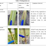 |
Table 3: Pathogenicity test |
Discussions
Identification of pathogenic fungal strains was carried out based on phenotypic and molecular characteristics. During this work, several species of Fusarium were identified. Their presence explains their involvement in the “Pokkah Boeng” disease. Indeed, the work of Hilton et al11 and those of Lin et al12 showed that several Fusarium species have been reported as causative agents of “Pokkah Boeng”, including F. sacchari, F. fujikuroi, F. verticillioides, F. andiyazi, F. proliferatum and F. moniliforme. The identification of Fusarium verticillioides on canes in the locality of Zuénoula would implicate it in the transmission of red snot. The samples collected in this locality showed red spots. Thus, Suresh and Nelson13 showed that the causative agent of red snot was Fusarium verticillioides. These results agree more or less with those of Kouadia et al14 who highlighted a diversity of phytopathogenic fungi belonging to the genera Colletotrichum, Fusarium, Aspergillus, Phoma, Penicillium, Curvularia, Botryodiplodia and Rhizoctonia associated with spoilage of banana, avocado and mango fruits. Furthermore, the genera Fusarium, Aspergillus, Botryodiplodia and Phoma have been reported on avocado and mango fruits15. These fungi are known to cause the deterioration of many products such as fruits, seeds, vegetables and tubers both in the field and during storage16. The fungal diversity recorded could be explained by the fact that sugar cane is a food with high nutritional value, mainly consisting of carbohydrates, water, vitamins and minerals. Also, the pH of sugar cane also being lower than 4.6, would more favor the development of pathogenic fungi17. Naturally, sugar cane resists microbial attacks, the extent of which will depend on the stage of maturity. This is because sugar cane contains high concentrations of natural antimicrobial compounds. These compounds consist of mixtures of 5-substituted resorcinols such as resorcinol-5-(12-heptadecadienyl) and resorcinol-5-(pentadecyl) Prusky et al18 accumulate in the stem. Faced with such a chemically unfavorable environment, the fungus suspends its development until the concentration of antimicrobial compounds decreases in the stem. The results of pathogenicity tests carried out on the canes showed that all of these isolated fungi were pathogens of sugarcane. Schizophyllum commune was the species that caused the most serious pathologies of sugar cane in the two localities after the Mosaic. This could be explained by the fact that this species is involved in anthrax. These results are in agreement with those of Yao et al19 who showed that the pathology spread very quickly in all sugar cane producing countries causing numerous yield losses ranging from 30 to 50% for susceptible varieties.
Conclusion
This present study contributed to the identification of pathogenic fungal germs of sugar cane in Côte d’Ivoire. From this study, it should be noted that the fungal species, namely Fusarium verticillioides, Fusarium subglutinans and Schizophyllum commune, were identified as pathogens of sugar cane in Côte d’Ivoire. These fungi are believed to be involved in pathologies such as red snot, “pokkah boeng” and smut, which are commonly encountered in sugar cane plantations.
Acknowledgment
The authors are thankful for the sugar society SUCRIVOIRE of Côte d’Ivoire.
Funding Sources
The author(s) received no financial support for the research, authorship, and/or publication of this article.
Conflict of Interest
The authors do not have any conflict of interest.
Data Availability Statement
This statement does not apply to this article.
Ethics Statement
This research did not involve human participants, animal subjects, or any material that requires ethical approval.
Informed Consent Statement
This study did not involve human participants, and therefore, informed consent was not required.
Clinical Trial Registration
This research does not involve any clinical trials.
Permission to reproduce material from other sources
Not Applicable
Author Contributions
Djédji Okoue, Armist Amoa Tehua and Ibrahima Kebe : Performed the majority of the experiments and data analysis.
Mireille Waze Alloue-Boraud : Designed the experiments and analysed the data.
Fulgence Yao Koffi : Wrote the manuscript.
All authors read and approved the final version of the manuscript.
References
- Péné B.C, Souleymane N, Chantal N. K. Sprinkler irrigation and soil tillage practices in sugarcane plantations as influenced by soil texture and water storage in northern Ivory Coast. J. Appl. Biosci. 2012;54:3916-3924.
- Kouamé D. K, Péné C. B, Zouzou M. Évaluation de la résistance variétale de la canne à sucre au foreur de tiges tropical africain (Eldana saccharina Walker) en Côte d’Ivoire. J. Appl. Biosci. 2010;26:1614-1622.
- Cawoy H, Bettiol W, Fickers P, Ongena M. Bacillus-Based Biological Control of Plant Diseases, Pesticides in the Modern World – Pesticides Use and Management, Dr. Margarita Stoytcheva (Ed.), ISBN: 978-953-307-459-7, InTech, 2011. Available from: http://www.intechopen.com/books/ pesticides-in-the-modern-world-pesticides-use-and-management/bacillus-based-biological-control-of-plant-diseases
CrossRef - Silué N, Soro K, Koné T, Abo K, Koné M, Koné D. Parasitical fungi in cashew ( Anacardium occidentale L.) orchard of Cote d’Ivoire. Plant Pathol. J. 2017;16:82-88. DOI: 10.3923/ppj.2017.82.88
CrossRef - Botton B., Breton A., Fevre M., Gauthier S., Guy P. H, Larpent J. P, Reymond P, Sarglier J. J, Vayssier Y, Veau P. (ed) : Usefull moist and nusible: industrial importance , 2nd edn Paris. Milan. Barcelone : Masson. « Collection Biotechnologies (Paris) ».1990;52 p.
- Guiraud J. P. (ed) : Microbiologie alimentaire, Paris : Dunod, (1998),652p.
- Maymonna S.N. Recherche de diversité génétique au sein d’une population Stemphylium solani Weber au Sénégal. Rapport de stage. 1998 : 66 p.
- Bessadat N., Simoneau P., Benichou S., Setti B., Kihal M, Henni D. E. Morphological, physiological and pathogenic variability of small-spore Alternaria sp. causing leaf blight of Solanaceous plants in Algeria. Afr.J. Microbiol. Res.2014 ;8(37):3422-3434. https://doi.org/10.5897/AJMR2014.6802
CrossRef - Hall T.A. BioEdit: A User-Friendly Biological Sequence Alignment Editor and Analysis Program for Windows 95/98/NT. Nucleic Acids Symp. Ser.1999;41:95-98.
- Silué N, Abo K, Johnson F, Camara B, Koné M, Koné D. Evaluation in vitro et in vivo de trois fongicides de synthèse et d’un fongicide biologique sur la croissance et la sévérité de collectotrichum gloesporioides et pestalotia heterocornis, champignons responsables de maladies foliaires de l’anacardier (Anacardium occidentale L.) en Cote d’Ivoire. Agron. Afr. 2018;30:107-122.
- Hilton A, Zhange H, Yu W, Shim B. W. Identification and Characterization of Pathogenic and Endophytic Fungal Species Associated with Pokkah Boeng Disease of Sugarcane. Plant Pathol. J. 2017;33(3): 238-248. DOI: 10.5423/PPJ.OA.02.2017.0029.
CrossRef - Lin Z, Xu S, Que Y, Wang J, Comstock J. C, Wei J, Zhang M. Species-specific detection and identification of Fusarium species complex, the causal agent of sugarcane pokkah boeng in China. PloS One. 2014;9(8):104195. DOI: 10.1371/journal.pone.0104195.
CrossRef - Suresh N, Nelson R. Diversity of Arbuscular Mycorrhizal fungi (AMF) in the rhizosphere of sugarcane. Eur. J. Exp. Biol.2016;5(3):13-19.
- Kouadia A.M-J, Abo K , Kouadio K.T. Evolution des infections naturelles sur les mangues, les avocats et les bananes en Côte d’Ivoire et principaux champignons reponables. J. Appl. Biosci.2019;134:13710-13721. DOI:10.4314/jab.v134i1.8.
CrossRef - Djeugap F.J, Tsompbeng G.N, Keuete K.E, Yaouba A, Serferbe S. Isolation and Identification of Fungi Associated with Avocado fruits from Local markets of the West Region of Cameroon. Inter J Agri Biosc. 2015;4(2):64-68.
- Dongmo G.Z, Djeugap F.J, Fenohi N, Kenfack N.D, Takuete R, Teguefouet P. Contribution à l’identification des champignons de post-récolte associés aux amandes de Ricinodendron heudelotii et Garcinia kola collectées dans les Hauts Plateaux de l’Ouest Cameroun. Int. J. Biol. Chem. Sci. 2017 ;11(4) :1840-1850.DOI : http://dx.doi.org/10.4314/ijbcs.v11i4.33
CrossRef - Cissé M. .Immobilisation d’un système lactoperoxyde dans un enrobage de chitosane dans le but de prolonger la conservation des mangues. Thèse de doctorat. Université Montpellier Supagro, France. 2012:159p. Cirad – Agritrop (https://agritrop.cirad.fr/566848/).
- Prusky D, Kobiler I, Miyara I, Alkan N. Fruit diseases” The Mango: Botany, Production and Uses. 2009:210-230. https://doi.org/10.1079/9781845934897.0210.
CrossRef - Yao K. J-E, Kouamé K. D, Kouamé K. G, Kassi K. F. J-M, N’guessan A. C, Eponon E. C. G, Koné D. Structure of Sporisorium scitamineum isolates, the causative agent of sugarcane smut in Côte D’ivoire. IJSR. 2020; 9(3):1376-1381. DOI: 10.21275/SR20307072052.

This work is licensed under a Creative Commons Attribution 4.0 International License.


